Bioengineering artificial microfluidic habitats for microbial single-cell analysis

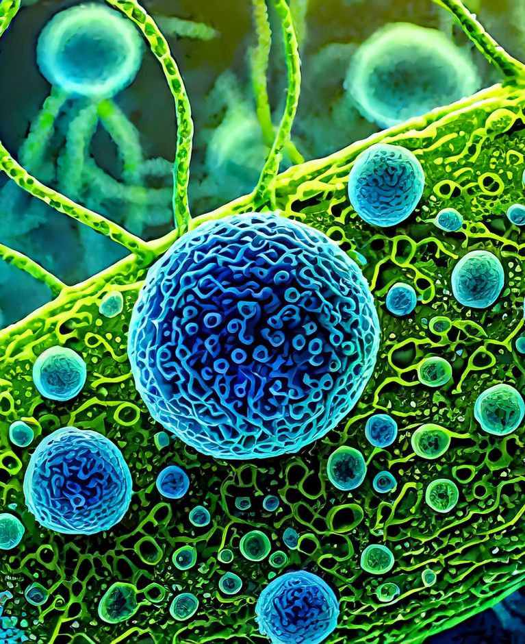
Figure 1 Image showing a microfluidic cultivation chip. A single chip incorporates up to 1000 micro chambers for culturing microorganisms under precise micro environmental control. Cell-cell and cell-environmental interactions can be followed by time-lapse microscopy.
Microorganisms, which include bacteria, viruses, fungi and archaea, are fundamental to life on Earth and play essential roles in diverse ecosystems as well as in human health. However, our current understanding of microbial functionality and the amazingly complex interactions within microbiomes is still rudimentary, as it is usually derived from model organisms studied under simple environmental control, poorly reflecting natural conditions. However, ever-advancing microfabrication technologies, which allow the creation of synthetic microfluidic environments, and advanced live-cell microscopy have the potential to revolutionize experimental microbiology by unlocking unseen spatially and temporally resolved microbial behavior and cellular interactions.
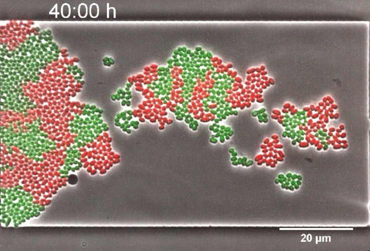
Figure 2 Life is more efficient with the help of others. This is also true for microorganisms. Time-lapse microscopy image of a synthetic microbial co-culture growing in a microfluidic chamber. The population consists of two genetically engineered strains (red and green fluorescing) of Corynebacterium glutamicum. This mixed culture can only grow in the presence of both strains simultaneously, as each strain provides an essential amino acid for which its interaction partner is auxotrophic.
IBG-1 at Forschungszentrum Jülich has been successfully pioneering the development of microfluidics for applied and basic research in microbiology and biotechnology since 2010. Innovative bottom-up concepts for microbial single-cell analysis enable researchers to address questions that cannot be answered by conventional laboratory-scale approaches. In highly interdisciplinary projects, microstructures at the femto- to picoliter scale are used to grow microorganisms with diverse requirements, providing new insights into mixed cultures, population heterogeneity, cellular interactions, single-cell physiology and more. Fluids behave differently at the micrometer scale. Laminar flow and diffusion governed mass transport can be precisely controlled, allowing precise spatial and temporal control of the microenvironment. Recent focus has been on advanced chip functionality and the development of more complex microfluidic landscapes to mimic natural, structured and dynamic environments. Deep learning algorithms are also being developed to efficiently analyze large image data stacks obtained by automated live-cell microscopy. The resulting software tools now enable nearly automated cell segmentation, cell tracking in dense populations, and discrimination between different organisms and fluorescently labelled strains.
More details:
Publications:
Enabling oxygen-controlled microfluidic cultures for spatiotemporal microbial single-cell analysis. Keitaro Kasahara, Markus Leygeber, Johannes Seiffarth, Karina Ruzaeva, Thomas Drepper, Katharina Nöh, Dietrich Kohlheyer. Front. Microbiol., 20 June 2023 Sec. Systems Microbiology Volume 14 - 2023 https://doi.org/10.3389/fmicb.2023.1198170
Assessing the growth kinetics and stoichiometry of Escherichia coli at the single-cell level. Katharina Smaluch, Bastian Wollenhaupt, Heiko Steinhoff, Dietrich Kohlheyer, Alexander Grünberger, Christian Dusny. Eng Life Sci. 2022 May 6;23(1):e2100157. https://doi.org/10.1002/elsc.202100157. eCollection 2023 Jan.
Microbial single-cell analysis in picoliter-sized batch cultivation chambers. Eugen Kaganovitch, Xenia Steurer, Deniz Dogan, Christopher Probst, Wolfgang Wiechert , Dietrich Kohlheyer. .N Biotechnol. 2018 Dec 25:47:50-59. https://doi.org/10.1016/j.nbt.2018.01.009 Epub 2018 Mar 14.
Spatiotemporal microbial single-cell analysis using a high-throughput microfluidics cultivation platform. Alexander Grünberger, Christopher Probst, Stefan Helfrich, Arun Nanda, Birgit Stute, Wolfgang Wiechert, Eric von Lieres, Katharina Nöh, Julia Frunzke, Dietrich Kohlheyer. Cytometry A . 2015 Dec;87(12):1101-15. https://doi.org/10.1002/cyto.a.22779 Epub 2015 Sep 8.
Contact
Dietrich Kohlheyer
Forschungszentrum Jülich
E-Mail